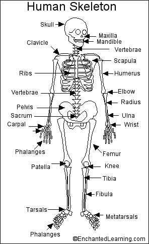
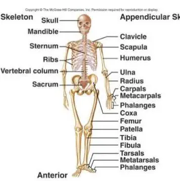
In the diaphyses of long bones, the bone marrow is yellow and contains predominantly fat, along with some marrow cells and some connective tissue. The hyoid is a U-shaped bone found at the base of the jaw. Typically, the spine follows gentle forward and backward curves. skeleton human bones printout enchantedlearning diagram labeled label body labelled skeletal class system Systemic pathologies that can weaken bone vary from osteoporosis (a decrease in bone mass per unit volume), osteomalacia (a decrease in the mineralization of the osteoid, occurring because of nutritional deficiencies, an adult variant of rickets), and hyperparathyroidism, among others. anatomy quiz skeleton axial bones skeletal system human appendicular worksheet body skull diagram test pdf labels answers multiple questions activities The appendicular skeleton comprises the bones of the extremities. At birth, the skeleton of a newborn has more than 300 bones; as a person ages, these bones grow together and fuse into larger bones, leaving adults with only 206 bones. Each finger has three bones known as phalanges, except for the thumb, which only has two phalanges. axial skeletal pe divisions labeling This is called fat embolism. The periosteum is attached to bone by fine fibers called Sharpey fibers. Cancer can develop in the tissues of the bone or in the cells produced by bones. Healthline Media does not provide medical advice, diagnosis, or treatment. The ulna is on the medial side of the forearm and forms a hinge joint with the humerus at the elbow.  The regions of each bone where muscles attach to the bone grow larger and stronger to support the additional force of the muscle. It is also the tissue from which most bones develop in children. These apertures are found more often at the metaphyseal ends of the long bones. Shoulder: highly mobile ball-and-socket joint with multiaxial movements. They connect to the subperiosteal space via Volkmann canals and to each other via canaliculi. Such a picture can be seen with long-standing anterior cruciate ligament tears in the knee or following meniscectomy. The calcified areas spread out from their blood vessels replacing the old tissues until they reach the border of another bony area. Proper levels of calcium ions in the blood are essential to the proper function of the nervous and muscular systems. Roughly half of the bone matrixs mass is water, while the other half is collagen protein and solid crystals of calcium carbonate and calcium phosphate. Human Osteology and Skeletal Radiology: An Atlas and Guide. They also serve as attachment points for various ligaments. skeletal diagram bones ecdn Some of these are the proximal tibial anatomical plate, anatomical plates for fixation of the distal humerus, proximal humeral anatomical locking plates (PHILOS), mini-fragment plates for small long bones, and others, providing extensive options for the management of specific injuries. Its made up of the clavicle (collarbone) and scapula (shoulder blade). There are 3 types of bone tissue, including the following: Compact tissue. The skeleton acts as a scaffold by providing support and protection for the soft tissues that make up the rest of the body. Finally, red bone marrow stores some iron in the form of the molecule ferritin and uses this iron to form hemoglobin in red blood cells. The bones of the superior portion of the skull are known as the cranium and protect the brain from damage. The tarsals form joints with the five long metatarsals of the foot. The skeleton begins to form early in fetal development as a flexible skeleton made of hyaline cartilage and dense irregular fibrous connective tissue. The bony skeleton is divided into 2 parts: the axial skeleton and the appendicular skeleton. Proximal Femoral Focal Deficiency/Congenital Femoral Deficiency, One Bout of Resistance Training Does Not Enhance Metformin Actions in Prediabetic and Diabetic Individuals. Cartilage is the specialized, gristly connective tissue that is present in adults. diagram anatomy skeleton human anatomical chart These offshoots are specific to each bone, depending on the bone's relations with its surrounding soft tissues. Long bones follow the process of endochondral ossification where the diaphysis grows inside of cartilage from a primary ossification center until it forms most of the bone. body skeleton galen anatomy history middle illustrations ages galens class bones medieval history13 stanford edu web shape discovered he In addition, there may be articular injuries to the ligaments and cartilage that further compromise the joint. Articular injuries are different from diaphyseal injuries. The muscle begins at the flexor retinaculum in, The movement of the upper arm and shoulder is controlled by a group of four muscles that make up the rotator cuff. The Latin translation of 'quadriceps' is 'four headed,' as the group, The palmaris brevis muscle lies just underneath the skin. The multiaxial shoulder joint and the uniaxial elbow joint allow the forearm and hand to be positioned for optimal function. The skeletal systems primary function is to form a solid framework that supports and protects the bodys organs and anchors the skeletal muscles. Early range of motion after accurate anatomical alignment and stable fixation of intraarticular fractures allows the cartilage to heal without defects and prevents the formation of intraarticular and extraarticular adhesions, thus minimizing disabilities. Once the long bone parts have fused together, the only hyaline cartilage left in the bone is found as articular cartilage on the ends of the bone that form joints with other bones. There are 206 bones in the human skeleton, not including teeth and sesamoid bones (small bones found within cartilage): 80 axial bones. Synovial joints are lined by a specialized membrane called the synovial membrane and the articulating ends of the bones are lined by specialized cartilage termed hyaline cartilage. The medullary cavity contains red bone marrow during childhood, eventually turning into yellow bone marrow after puberty. The periosteum contains blood vessels, nerve fibers, osteoblasts, and osteoclasts. human organs body diagram vector The human skeletal system consists of all of the bones, cartilage, tendons, and ligaments in the body. There are also some differences in the male and female skeleton. These tissues act as a soft, growing framework and placeholder for the bony skeleton that will replace them. Anatomically and structurally, the different types of bone are traditionally grouped as follows: Long bones - Clavicle, humerus, radius, ulna, metacarpals, femur, tibia and fibula, metatarsals, and phalanges; the metacarpals, metatarsals, and phalanges are sometimes referred to as short long bones, Flat bones - Skull, mandible, scapula, sternum, and ribs, Short bones - Carpal and tarsal bones, patella, and sesamoids Irregular bones - Vertebrae, sacrum, coccyx, and hyoid bone. The axial skeleton runs along the bodys midline axis and is made up of 80 bones in the following regions: The appendicular skeleton is made up of 126 bones in the folowing regions: The skull is composed of 22 bones that are fused together except for the mandible. This article provides an overview of the basic anatomy of the human skeleton, bones, and joints (see the image below). Formed by the left and right hip bones, the pelvic girdle connects the lower limb (leg) bones to the axial skeleton. Matsches E, Burbridge B, Sher B, Mohamed A, Juurlink B. The skeletal system also provides attachment points for muscles to allow movements at the joints. This function is especially evident with subcutaneous bones like the tibia: when such bones are exposed and injured, their very survival may depend on the presence or absence of periosteum. A small band of hyaline cartilage remains in between the bones as a growth plate. Fat cells are also found within the bone marrow. The normal anatomy of bone is exploited in the management of various injuries of bone. Red bone marrow produces red and white blood cells in a process known as hematopoiesis. Bone injuries invariably damage the soft tissue in the vicinity to variable extents. The smooth tissue at the ends of bones, which is covered with another type of tissue called cartilage. Mayo Clinic Staff. The pectoral girdle connects the upper limb (arm) bones to the axial skeleton and consists of the left and right clavicles and left and right scapulae. www.bartleby.com. Regardless of age or sex, the skeletal system can be broken down into two parts, known as the axial skeleton and the appendicular skeleton. Altogether, the skeleton makes up about 20 percent of a persons body weight. The vertebral column is made up 26 bones. Almost every skeletal muscle works by pulling two or more bones either closer together or further apart. The organic phase is formed by cells and the collagen-forming part of the matrix. If you log out, you will be required to enter your username and password the next time you visit. The skeleton makes up about 30-40% of an adults body mass. The male skeleton is usually longer and has a high bone mass. [2] The cellular content of the red marrow consists primarily of rounded nucleated cellsthe true marrow cells, or, as Gray calls them, the marrow cells of Kolliker (or myelocytes). The yellow bone marrow inside of our hollow long bones is used to store energy in the form of lipids. Innerbody Research does not provide medical advice, diagnosis, or treatment. The tibia and fibula form the ankle joint with the talus, one of the seven tarsal bones in the foot. The surfaces of long bones and flat bones have ridges and surfaces that are formed by the attachments of muscles and ligaments. Bone is a solid organ that appears pinkish white externally and deep red internally when in a fresh state. View of human skeleton from behind, showing rib cage and spine. Accessed: April 2011. Masks are required inside all of our care facilities. Pay attention to joint pain and any changes you perceive in your ability to move, sharing those with your healthcare provider. The vast difference in height and limb length between birth and adulthood are mainly the result of endochondral ossification in the long bones. Stem cells and osteoblast cells in the periosteum are involved in the growth and repair of the outside of the bone due to stress and injury. These lamellae are often called by different names, depending on their location; they may be interstitial lamellae, primary or fundamental lamellae, or circumferential lamellae. anatomy body female upper abdomen illustration diagram istockphoto The masseter is the primary muscle that brings your teeth together when youre chewing. Living bone cells are found on the edges of bones and in small cavities inside of the bone matrix. Fibrous joints exist where bones are very tightly joined and offer little to no movement between the bones. 2008 Nov. 3 Suppl 3:S131-9. skeleton bones dinosaur human lesson study skeletons Examples of synovial joints include the knee, hip, elbow, and atlanto-axial joint.
The skeletal system in an adult body is made up of 206 individual bones. This fluid also nourishes the articular cartilage, which has sparse blood supply. An adults skeleton contains 206 bones. It is therefore imperative that articular and juxtaarticular injuries be treated with modalities that allow early restoration of joint motion and early rehabilitation. anatomical diagrams drawings figure (2009). (2016). Long bones have a spongy bone on their ends but have a hollow medullary cavity in the middle of the diaphysis. skeleton skeletal teachpe physiology labelled
The tibia and fibula are the bones of the lower leg. Twenty-six vertebrae form the vertebral column of the human body. We are vaccinating all eligible patients. The direction and intensity of force determines the degree of damage to bone. organs abdomen quadrant quadrants digestive location boundaries diaphragm The femur forms the ball and socket hip joint with the hip bone and forms the knee joint with the tibia and patella. Some diaphyseal injuries like those in the humerus and the radius and ulna are best managed by fixation with plates and screws as these allow speedy restoration of function with minimum postoperative immobilization. This precludes most nonsurgical methods of treatment. Cancers of the blood cells produced by bone, such as myeloma or lymphoma, are more common. The bones of the inferior and anterior portion of the skull are known as facial bones and support the eyes, nose, and mouth. Images depicting the anatomy of the adult skeleton can be seen below. The tibia is much larger than the fibula and bears almost all of the bodys weight. This makes the joint specially suited for mobility. Cancellous bone, on the other hand, is composed of bony trabeculae that run along the lines of stress. Ligament injuries to weight-bearing joints have a similar impact on the articular cartilage. Blood vessels present in the periosteum provide energy to the cells on the surface of the bone and penetrate into the bone itself to nourish the cells inside of the bone. The supraspinatus muscle is a rotator cuff muscle located in the shoulder, specifically in the supraspinatus fossa, a concave depression in the rear, The quadratus plantae is a muscle in the foot that extends from the anterior (front) of the calcaneus (heel bone) to the tendons of the digitorum. Bone is covered by a membrane called the periosteum. The structural peculiarities of the human skeleton give human beings their characteristic appearance and facial geometry. Learn more: Vaccines, Boosters & Additional Doses | Testing | Patient Care | Visitor Guidelines | Coronavirus | Email Alerts. They can be due to things such as a deficiency in vitamin D, loss of bone mass, and use of certain medications, such as steroids or chemotherapy. Arthritis is an inflammation of the joints. Normal bone anatomy and physiology.
New blood cells are produced by the red bone marrow inside of our bones. Also, you can learn more about DNA health tests, which can tell you if youre at a genetically higher risk of hemochromatosisone of the most common hereditary disorders, causing joint painas well as Gaucher disease. It is a short muscle on the flat of the hand. Osteoclast. These bones are arranged into two major divisions: the axial skeleton and the appendicular skeleton. In addition, the overall mass and thickness of a bone increase when it is under a lot of stress from lifting weights or supporting body weight. Haversian canals run longitudinally down the bone. Each toe has three phalanges, except for the big toe, which only has two phalanges. Joint injuries are also associated with a potential for stiffness and disability consequent to the loss of range of motion. The 24 vertebrae can be further divided into the: The sacrum and coccyx are both made up of several fused vertebrae. Bones often act as levers, which, in conjunction with muscular contraction, initiate and sustain movement. Males have larger skeletal size and bone mass than females, despite comparable body size. system skeletal anatomy physiology The bones of the skeletal system act as attachment points for the skeletal muscles of the body. Some of the ribs attach directly to the sternum, while others are linked to the sternum via cartilage. The hyoid is the only bone in the body that does not form a joint with any other boneit is a floating bone. The hyoid is a small, U-shaped bone found just inferior to the mandible. The periosteum is a vital structure in bone function, serving to nourish and protect the underlying cortical bone. Children tend to have more red bone marrow compared to their body size than adults do, due to their bodys constant growth and development. (n.d.). Fractures typically occur due to an injury or trauma, such as a car accident or a fall. The thoracic cage is made up of the sternum (breastbone) and 12 pairs of ribs. Subchondral tissue. It consists of the bones that make up the arms and legs, as well as the bones that attach them to the axial skeleton. As we grow through childhood, the growth plates grow under the influence of growth and sex hormones, slowly separating the bones. Metabolic bone diseases refer to a group of conditions that affect bone strength or integrity. There are three main types of spinal curvature: The skeletal system provides the foundation for all of the bodys movements, in addition to other important functions. These are lined by articular or hyaline cartilage. hand bones bone diagram anatomy hands left In the skull these soft spots are known as fontanels, and give the skull flexibility and room for the bones to grow. organs organ labeled etymologies woldia etymology reproductive bone gland massive cells exatin healthiack The skeletal system includes all of the bones and joints in the body. femur bone diagram leg anatomy human anterior 3d infographic system medical body (n.d.). The upper limbs are designed to enable sophisticated movement of the hand in space so as to serve the unique prehensile and fine activities that can be performed with the upper limb. Introduction to the skeletal system. These 21 fused bones are separate in children to allow the skull and brain to grow, but fuse to give added strength and protection as an adult. The different types of bone cells include the following: Osteoblast. The periosteum also contains nervous tissue and many nerve endings to give bone its sensitivity to pain when injured. The cervical spine is lordotic (concave anteriorly), and the thoracic spine has a reverse curvature and is kyphotic (convex anteriorly). These bones form a protective cage around the organs of the upper torso, including the heart and lungs. The adult skull comprises 22 bones. Beneath the hard outer shell of the periosteum are tunnels and canals through which blood and lymphatic vessels run to carry nourishment for the bone. Then each of the metatarsals forms a joint with one of the set of phalanges in the toes. Anterior view of lumbar spine, showing lumbar vertebrae, curvature of lumbar spine, sacroiliac joints, and iliac wings. The inorganic phase contains hydroxyapatite, calcium salts, and other minerals. A pathological fracture through a benign cyst in the proximal femur. The bone essentially then works as a reservoir of calcium, which is important for most metabolic functions in the human body. The inner medullary cavity is lined with a membrane called the endosteum. Our mission is to provide objective, science-based advice to help you make more informed choices. Synovial joints are the most common type of articulation and feature a small gap between the bones. Bone derives its blood supply from nutrient arteries, which enter the medullary canal at fixed points in the cortex. The amount of red bone marrow drops off at the end of puberty, replaced by yellow bone marrow. CRC Press; 2005. Nieves JW, et al. Intramedullary fixation is the standard of treatment of long bone diaphyseal injuries in the lower limb. Found in a small cavity inside of the temporal bone, they serve to transmit and amplify sound from the eardrum to the inner ear. Found within the bone, its function is to help maintain bone as living tissue. These fractures are treated differently from the usual traumatic fracture, the management being dictated by the pathology and its prognosis. The first 24 are all vertebrae, followed by the sacrum and coccyx (tailbone). At Another Johns Hopkins Member Hospital: Masks are required inside all of our care facilities, COVID-19 testing locations on Maryland.gov. The outside of a bone is covered in a thin layer of dense irregular connective tissue called the periosteum. Thomas R Gest, PhD is a member of the following medical societies: American Association of Clinical AnatomistsDisclosure: Nothing to disclose. In terms of the type of tissue that bridges them, joints may be described as synovial or fibrous, cartilaginous, or bony. 6.1: The functions of the skeletal system. Each arm contains 30 bones, known as the: The pelvic girdle, commonly known as the hips, is where the legs attach to the axial skeleton. This includes arms, shoulders, wrists, hands, legs, hips, ankles, and feet. This process continues until the end of puberty, when the growth plate stops growing and the bones fuse permanently into a single bone. Deep to the compact bone layer is a region of spongy bone where the bone tissue grows in thin columns called trabeculae with spaces for red bone marrow in between.